Anatomical and functional features of the knee joint
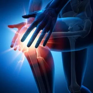
Knee joint — the largest joint in the human body. And although many people consider the hip joint to be the largest, in reality this is not the case. It is enough to compare X-rays of the hip and knee joints of one person, paying attention to the size of their articular surfaces. This misconception exists due to the fact that on the surface of the hip joint, unlike the knee joint, there is a pronounced layer of soft tissue, making it visually larger - developed muscles of the gluteal region and an extensive layer of subcutaneous fat.
The knee joint is in its structure complex(since it is formed by 3 bones: femur, tibia and patella) and complex(since it contains auxiliary cartilage - 2 meniscus). In shape, this joint is condylar (since the protruding part of the articular surface of the femur contains 2 condyles - medial and lateral), and in function - biaxial. Here, movements around 2 axes are possible - frontal (flexion and extension) and vertical (in a half-bent position, internal and external rotation).
There is a complex tendon-ligamentous apparatus, auxiliary cartilaginous formations (menisci), large folds of the synovial membrane, many synovial bursae, the articular cavity in the area of the patella forms a large superior inversion.
The knee joint bears a significant load, because with a vertical load it is located at the bottom of the body. The longest arms of the human skeleton—the femur and tibia—articulate at this joint. With a healthy, normally functioning knee joint, this entire complex system operates harmoniously and harmoniously, providing the function of support and movement of the lower limb in a wide variety of conditions, under the influence of both small and large loads.
Osteoarthritis of the knee joint
The complex structure, location in the human body and high functional requirements make the knee joint quite vulnerable. If any link of this system is affected, then the function of the entire knee joint and the lower limb as a whole immediately begins to suffer seriously.
Deforming osteoarthritis of the knee joint (gonarthrosis) is a chronic degenerative-dystrophic disease. It is based on the initial degeneration of articular cartilage with subsequent changes in the bone articular surfaces and the development of marginal osteophytes (bone spines), as well as the involvement of all elements of the joint in the pathological process. In the future, these changes lead to deformation of the joint, and sometimes to the development of moderate synovitis.
Arthrosis can be primary(idiopathic) - it develops in initially healthy articular cartilage under the influence of chronic overload of the joint. Orsecondary - develops even with normal joint loads due to preliminary pathological changes in the articular cartilage (due to injuries, chronic inflammation, circulatory disorders, endocrine and metabolic disorders).
Features of diagnosing arthrosis of the knee joint
Both primary and secondary arthrosis of the knee joint, affecting the articular cartilage and underlying bone tissue, can additionally involve a variety of joint structures in the pathological process. The fact is that bone and cartilage tissue do not have pain receptors, which means they cannot be a source of pain a priori. Therefore, the individual nature of joint pain (pain pattern) depends precisely on which anatomical structures are involved in the pathological process and on the number of these affected structures.
Sometimes arthrosis of the knee joint can be accompanied by synovitis (inflammation of the inner lining of the joint capsule with the production of inflammatory fluid and excessive production of synovial fluid), leading to excessive accumulation of fluid in the joint cavity. This further complicates treatment.
As a rule, in a knee joint affected by arthrosis, changes occur over time in almost all soft tissue structures. There is rigidity of the ligamentous apparatus and periarticular tendons, the menisci are fragmented, the fat body of the synovial fold is hyperplastic, the synovial bursae contain an increased volume of synovial fluid. But not all of these structures are the source of pain in each specific case. Therefore, the art of diagnosis and treatment lies in correctly identifying all the anatomical structures involved in the pathological process and directing efforts to cure them. This approach to treatment will be the most effective - This is an individual approach.
Only by analyzing an X-ray, tomogram, or ultrasound, it is impossible to reliably determine where exactly the pain in a particular patient is. With very similar images, different patients may experience pain in completely different places in the knee joint. Therefore, in such a situation, a medical clinical examination is of paramount importance. And here the experience of a doctor who sets himself the task of dealing with the problem of a particular patient is extremely important.
Features of the treatment of arthrosis of the knee joint
In advanced cases of arthrosis of the knee joint (III degree) traditional approach ineffective in treatment. The patient is faced with a limited choice: either undergo knee replacement surgery (endoprosthetics), or find a treatment method that will help without surgery.
During the operation the joint is completely removed (bone articular ends, degeneratively changed joint capsule, ligaments, menisci, etc.) and an endoprosthesis is installed in their place. With effective non-surgical treatment all structures of the knee joint, including those that are the source of pain, are preserved, but thanks to the therapeutic effect directed at them, the inflammatory process in them stops and the pain goes away along with it. As a result, function improves and the sick person feels healthy again.
This non-surgical treatment is used in theArthroklinik Akta®, demonstrating its effectiveness for a number of years.
Treatment of arthrosis of the knee joint at Arthroclinic Akta®
The essence of this technique is injection treatment of anatomical structures responsible for pain in this particular case. The treatment technique used does not place emphasis on the use of scarce potent drugs. Of primary importance is the choice of injection sites and technique for their implementation.
During the treatment procedure, a series of injections are made into certain non-standard places (the injection sites are individual in each specific case). In this case, injections are made without penetrating into the joints and spinal canal, as well as without penetrating any “dangerous” places at all.