Currently, more and more often, with diseases of the musculoskeletal system (especially with pathology of the spine), patients are referred for tomographic examination, completely ignoring the good old X-ray method. How justified is this approach? After all, it is doctors who often tell the patient: “An X-ray won’t show anything, you need to do a tomogram.”
In the vast majority of cases, additional studies need to begin with an x-ray.
It is irrational to do a tomogram and skip an x-ray examination!
To answer this question, you need to understand what each method represents, as well as what data it can provide for making a diagnosis and choosing subsequent treatment tactics.
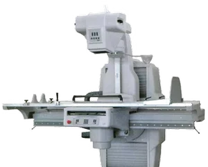
X-ray examination
Radiography is a method of radiation diagnostics that consists of obtaining a summary projection image of the anatomical structures of the body on X-ray film by passing X-rays through them and recording the degree of their attenuation.
Here it should be noted that the “old” X-ray method is relative. X-rays (X-rays) and x-ray diagnostic techniques were discovered by the German physicist Conrad Roentgen in 1895, for which he received the Nobel Prize in Physics in 1901.
Of the modern types of x-ray examination, special attention deservesdigital radiography(computer radiography) - a technology that allows you to obtain x-ray images of higher quality while minimizing the dose of x-ray radiation. Such images can be saved to digital media and subsequently subjected to additional processing on a computer (to optimize the brightness and contrast of the image).
Tomography
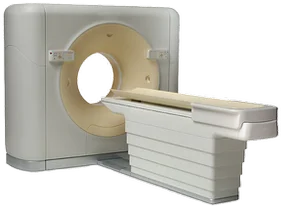
From ancient Greek. τομή - section. This is a radiation diagnostic method that allows you to obtain a layer-by-layer image of areas of the human body.
The method is based on the measurement and complex computer processing of the difference in the attenuation of X-ray radiation by tissues of different densities.
The most common are 2 types of tomography, differing in the type of radiation used. These are X-ray and magnetic resonance imaging.
Computed tomography (X-ray computed tomography, CT, RCT) is a tomographic method for studying the internal organs of a person using x-rays.
In a broad sense, “computed tomography” is a synonym for the term “tomography” (since all modern tomographic methods are implemented using computer technology). In a narrow sense (in which it is used much more often), it is a synonym for the term “X-ray computed tomography”, since it was this type of method that laid the foundation for modern tomography.
CT was proposed in 1972 by Godfrey Hounsfield and Allan Cormack, who were awarded the 1979 Nobel Prize in Physiology or Medicine for this development.
In 1986, the idea of spiral scanning of the area under study was invented and patented in Japan. In this case, the trajectory of the X-ray tube towards the longitudinal axis of the object under study takes on the shape of a spiral. Spiral scanning technology has made it possible to significantly reduce the time spent on CT examinations and significantly reduce the radiation dose to the patient.
In 1992, multislice computed tomography (“multislice”, “multi-slice” computed tomography, MSCT) was introduced - using devices with gantry apertures around the circumference of not one, but two or more rows of detectors. This further improved the quality of the study.
CT uses x-rays to provide information about the physical state of a substance. Moreover, the denser the fabric, the more intense the shadow it gives and the better visualized it is.
Magnetic resonance imaging (MRI, NMR imaging, MRI, NMR) is a non-radiological tomographic method for studying the internal organs of a person using a strong magnetic field. Based on the NMR (nuclear magnetic resonance) effect - measurement of the electromagnetic response of the nuclei of hydrogen atoms to their excitation by a certain combination of electromagnetic waves in a constant magnetic field of high intensity. The greater the fluid content in the tissue, the more intense the response of the nuclei of hydrogen atoms.
The year 1973 is considered to be the founding year of magnetic resonance imaging. Peter Mansfield and Paul Lauterbur received the Nobel Prize in Medicine in 2003 for their invention of MRI.
For some time, the term NMR imaging existed, which was replaced by MRI in 1986 due to the development of radiophobia in people after the Chernobyl accident. In the new term, the reference to the “nuclear” origin of the method disappeared.
MRI uses a constant magnetic field and radio frequency electromagnetic radiation from the device to record the distribution of protons, that is, it gives an idea of the chemical structure of tissues.
Clinical significance of radiographs, CT and MRI
X-ray, unlike a tomogram, gives a better overview of the area under study.
Tomogram (CT and MRI), unlike a conventional x-ray, gives a better targeted view of the area of interest, and also at the required depth.
That is, it’s the same as when studying geography, looking at the globe as a whole and at maps of individual countries.
X-ray examination (conventional x-ray or computed tomogram) clearly visualizes dense tissue (bones, stones). But soft tissues are poorly identified.
Magnetic resonance imaging well visualizes soft tissues (muscles, brain, nerves, intervertebral discs, ligaments, etc.). But at the same time, dense tissues are less well defined.
Resume:
1. X-ray and tomogram are different and complementary studies. A tomogram is no better than an x-ray and in no case replaces it. She complements it.
2. In the vast majority of cases, the study should begin with a regular x-ray.This gives a good overview of the area under study and, in most cases, allows the correct diagnosis to be made already at this stage. In this case, there is no need for further referral for tomography.
In any process of cognition one should almost always go from the general to the specific. Those. It is wrong to try to understand the details without knowing the big picture.
It is irrational to do a tomogram right away, skipping an X-ray examination!
3. Digital x-ray is the best option for x-ray examinationBecause Compared to a conventional X-ray, the digital technique allows for better visualization of the area under study, and also has a number of other advantages.
4. If it is necessary to clarify the diagnosis, a tomographic examination may be prescribed after radiography and based on its data. In this case, in the direction of the tomogram, you need to indicate which parts of the study area need to be visualized especially carefully.
5. The choice of tomography typedepends on which tissues require more thorough examination. To study the bones of the skeleton, CT is prescribed, and to study soft tissues, MRI is prescribed.
In some cases, to clarify the diagnosis, it may be advisable to perform all types of studies - radiography, CT and MRI.